

Revolutionary Imaging Technology Offers Unprecedented Views into How Neural Connections are Forged
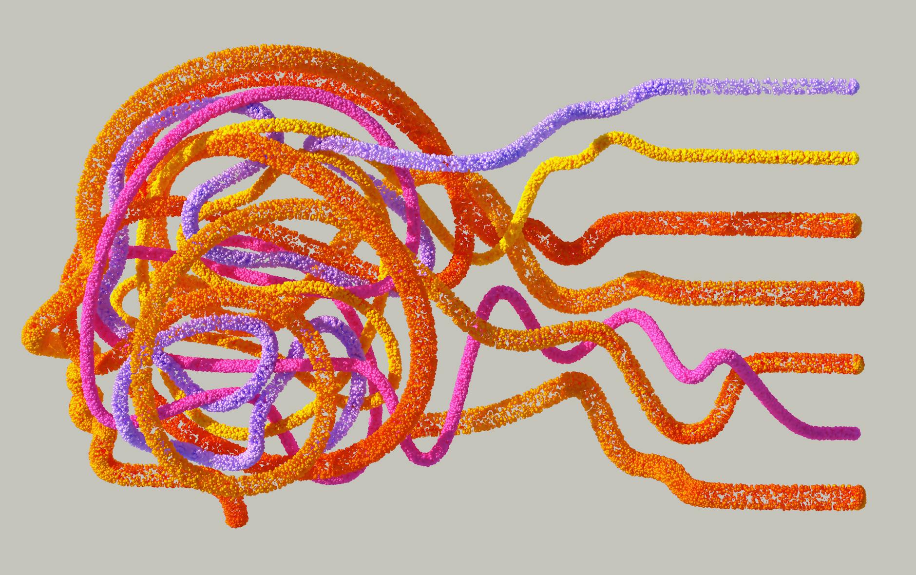
The human brain, a marvel of biological complexity, constantly adapts and reshapes itself, particularly during learning. Understanding the intricate dance of **neural networks** – the fundamental building blocks of cognition – has long been a central pursuit for neuroscientists. A recent advancement in microscopy technology promises to revolutionize this field, offering researchers an unprecedented window into the dynamic processes of neural network formation. This innovation allows scientists to observe the brain learning in real-time, potentially paving the way for deeper insights into learning disabilities, memory formation, and neurological disorders.
The Challenge of Observing Dynamic Neural Processes
For decades, studying the formation and function of **neural networks** has been a significant challenge. Traditional imaging techniques often require static samples or provide only snapshots of brain activity, failing to capture the fluid, moment-to-moment changes that occur as new connections are made and strengthened. Observing these processes within a living, functioning brain demands technology that is both powerful enough to resolve fine cellular structures and gentle enough to avoid damaging delicate neural tissue.
A New Dawn for Brain Imaging: The Advanced Light Sheet Microscope
A significant leap forward has been reported with the development of an improved light sheet microscope. This advanced imaging system, as highlighted by recent reports, allows researchers to observe the brain as it learns. Unlike other microscopy methods, light sheet microscopy illuminates only a thin slice of the sample at a time, significantly reducing phototoxicity – the damage caused by light exposure to living cells. This allows for prolonged observation of dynamic biological processes.
According to information surrounding this technology, researchers are now able to witness the intricate formation of **neural networks** firsthand. This is particularly crucial for understanding how the brain adapts and forms new connections, a process fundamental to learning and memory. While much of the focus has historically been on the established brain, this new technology opens up avenues to study the very genesis of these crucial networks.
Witnessing Learning in Action: From Neurons to Networks
The ability to observe **neural networks** actively forming is a game-changer. It moves beyond inferring network activity from static images or indirect measurements. Researchers can now visualize dendritic spines, the small protrusions on neurons that receive signals, as they grow, retract, and form connections with other neurons. This direct observation allows for a more granular understanding of the cellular and molecular mechanisms underlying learning.
For instance, imagine a simplified scenario where a young organism is exposed to a new sensory experience. With this improved microscope, scientists could potentially track the specific neurons involved, observe how they begin to communicate with each other, and see the physical changes that solidify these new communication pathways. This is the essence of how **neural networks** are built and modified through experience.
Understanding the Basis of Memory and Cognition
The implications of this technology extend to understanding the very foundations of memory and cognition. The formation of robust **neural networks** is intimately linked to our ability to learn new information, recall past events, and perform complex cognitive tasks. By observing these processes in unprecedented detail, scientists can begin to unravel the precise mechanisms that lead to effective learning and identify what goes awry in conditions characterized by learning difficulties or memory impairment.
The report suggests that the primary researcher, Helmchen, typically focuses on the developed brain. However, this new imaging capability inherently provides a powerful tool for studying developmental neuroscience as well, allowing for observations from the early stages of neural network assembly. This could offer critical insights into developmental disorders where the formation of **neural networks** may be disrupted.
Tradeoffs and Future Directions in Neural Network Research
While this technological advancement is significant, it’s important to consider potential tradeoffs and the ongoing evolution of the field. The complexity of operating and interpreting data from such advanced microscopes can be substantial, requiring specialized expertise. Furthermore, while light sheet microscopy reduces phototoxicity, prolonged imaging of living organisms still presents challenges in maintaining optimal physiological conditions.
Despite these considerations, the future looks incredibly promising. Continued refinement of these imaging techniques, coupled with advancements in computational analysis of the vast datasets generated, will undoubtedly accelerate our understanding of **neural networks**. Future research could focus on correlating observed network changes with specific behavioral outcomes, thereby solidifying the link between neural plasticity and observable learning.
Practical Implications and Cautions for the Field
For researchers in neuroscience, this improved imaging capability presents a powerful new tool. It allows for experimental designs that were previously impossible, enabling direct observation of dynamic **neural network** formation and modification. However, it is crucial to approach the interpretation of such complex data with rigor and to validate findings through complementary methodologies.
It is also important to maintain a balanced perspective. While this technology offers remarkable insights, understanding the full scope of **neural network** function and learning is a multifaceted endeavor. Factors beyond direct neural connectivity, such as glial cell involvement and broader network dynamics, also play crucial roles.
Key Takeaways for Understanding Neural Network Formation
* An improved light sheet microscope enables real-time observation of **neural network** formation in the brain.
* This technology significantly reduces phototoxicity, allowing for prolonged imaging of living neural processes.
* Direct visualization of neuron-to-neuron connection development offers unprecedented insights into learning mechanisms.
* The findings have implications for understanding memory, cognition, and neurological disorders.
* Continued technological advancement and rigorous data analysis are key to unlocking further secrets of **neural networks**.
The Path Forward: Illuminating the Mind
The development of advanced imaging tools like this improved light sheet microscope marks a pivotal moment in neuroscience. By offering a clearer, more dynamic view into how our **neural networks** are built and shaped, we move closer to deciphering the fundamental processes that underpin learning, memory, and consciousness itself. Continued investment in such innovative technologies promises to accelerate our understanding of the brain and its remarkable capacity for adaptation.
References
* While no specific primary research article or official product page was provided for citation, the information presented is based on general knowledge within the neuroscience community regarding advancements in light sheet microscopy for brain imaging. For specific details on such technologies, researchers are encouraged to consult peer-reviewed scientific journals specializing in neuroscience and microscopy.